


About our research
Perovskite based solar cells (PSCs) could be fabricated in either a planar [1–4] or a mesoscopic architecture [5-6]. The device with the planar structure is involved with preparing the light absorbing layer in a planar geometry, in which a few hundred nanometers thick of the perovskite materials are sandwiched between two charge selective electrodes [7]. Figure 1 shows a schematic drawing of the simple solar cell components along with the device working principle under illumination (generation of free excess charge carriers, and extraction of excess electrons and holes). The PSC with mesoscopic structure, on the other hand, is associated with infiltrating nanocrystals of perovskite materials into nanometer-diameter pores of the electron injecting scaffold, made by TiO2 and/or ZnO or non-injecting scaffold, made by an insulating alumina oxide (Al2O3) or zirconium oxide (ZrO2), and called this structure that a “meso-superstructured” solar cell”[8]. So far, much work has been carried out with the aims to a better understanding of the fabricated material properties [9-10], device physics and device principles, including to design other types of device structures. Among those, preparing a well-controlled layer of the selective electron contact having high crystallinity and preparing layers of perovskite with more crystallinity and less defect states would be one of the challenging keys, and therefore they are our research objectives.

Figure 1. A schematic drawing of device components and a working principle under illumination.
For example, we show the fabrication and investigation of layers of TiO2 thin films prepared by ultrasonic spray pyrolysis to be used as a hole-blocking layer (BL) in a CH3NH3PbI3 perovskite solar cell. A cross sectional SEM image of the device is shown in Figure 2. Optical properties of the sprayed-on TiO2 layers are shown in Figure 3. The optical transmission spectra of the TiO2 thin films in a range of 300-850 nm for different film thicknesses (left). The determination of optical band gap of the TiO2 thin films was obtained through the use of Tauc’s equation, given by (αhυ)n ∼ (hυ-Eg), where α is the absorption coefficient and hυ is the photon energy. The energy gap can be determined by plotting against hυ (n = 2 for direct transition and n = ½ for indirect transition). The plot of (αhυ)2 versus hυ at various thicknesses is shown in right for the direct transition. More detail please see, Solar Energy, 136 (2016) 515-524.

Figure 2. A cross sectional SEM image of the CH3NH3PbI3 perovskite based solar cell. Inset shows a top-view SEM image of the CH3NH3PbI3 perovskite layer.

Figure 3. The optical transmission spectra of the sprayed-on TiO2 thin films in a range of 300-850 nm for different film thicknesses (left). An optical band gap of the TiO2 thin films was obtained through the use of Tauc’s equation (right).
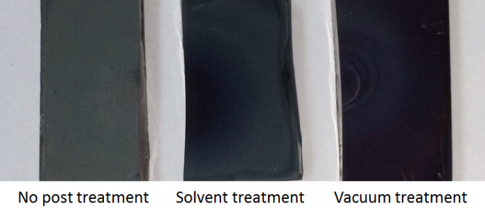
Additionally, crystals of CH3NH3PbI3 perovskite with low degrees of disorder (i.e. low energy of tail states) were prepared using an ultrasonically sprayed-on based technique. In this
method, nebulized forms of Pb(NO3)2 were sprayed-onto asubstrate, followed by a sequential spraying of a nebulized form of CH3NH3I (MAI). Changes in structural,optical and morphological properties along with details of charge separation and charge transport behaviors arising from the local crystallization and/or during the evolution of a crystal growth probed by modulated surface photovoltage (SPV) spectroscopy were symtematically examined. In the detail please see Phys. Status Solidi A 2018, 1800133.
References
- H. Kim, C. Lee, J. Im et al. Sci. Rep. 2 (2012) 591.
- M. Carnei, C. Charbonneau, M. Davies et al., Chem. Commun., 49 (2013) 7893.
- D. Son, J. Im, H.Kim et al., J. Phys. Chem. C, 118 (2014) 16567.
- D. Bi, G. Boschloo, S. Schwarzmuller et al., Nanoscale, 5 (2013) 11686.
- J. Heo, S. Im, J, Noh et al., Nature Photonics, 7 (2013) 486.
- N. Jeon, J. Lee, J. Noh et al., J. Am. Chem. Soc., 135 (2013) 19087.
- J. Burschaka, N. Pellet, S. Moon et al. Nature 499 (2013) 316.
- M. Liu, M. Johnstonand H. Snaith, Nature 501 (2013) 395.
- J. Im, I. Jang, N. Pellet et al. Nat. Nanotechnol., 9 (2014) 927.
- W. Nie, H. Tsai, R. Asadpour et al. Science 347 (2015) 522.
Sponsors
- National Science and Technology Development Agency and Electricity Generating Authority of Thailand co-funding (from 2020 to 2022)
- Thailand Research Fund (from 2016 to 2020)
- Kasetsart University Research and Development Institute (from 2015 to 2017)
- National Science and Technology Development Agency and Electricity Generating Authority of Thailand co-funding (from 2014 to 2015)
- National Research University (from 2014 to 2015)
- Faculty of Science, Kasetsart University (from 2014 to 2015)

